Speaker A
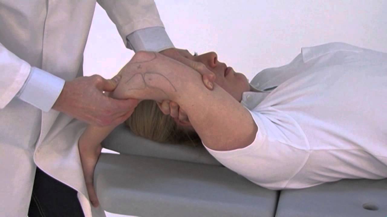
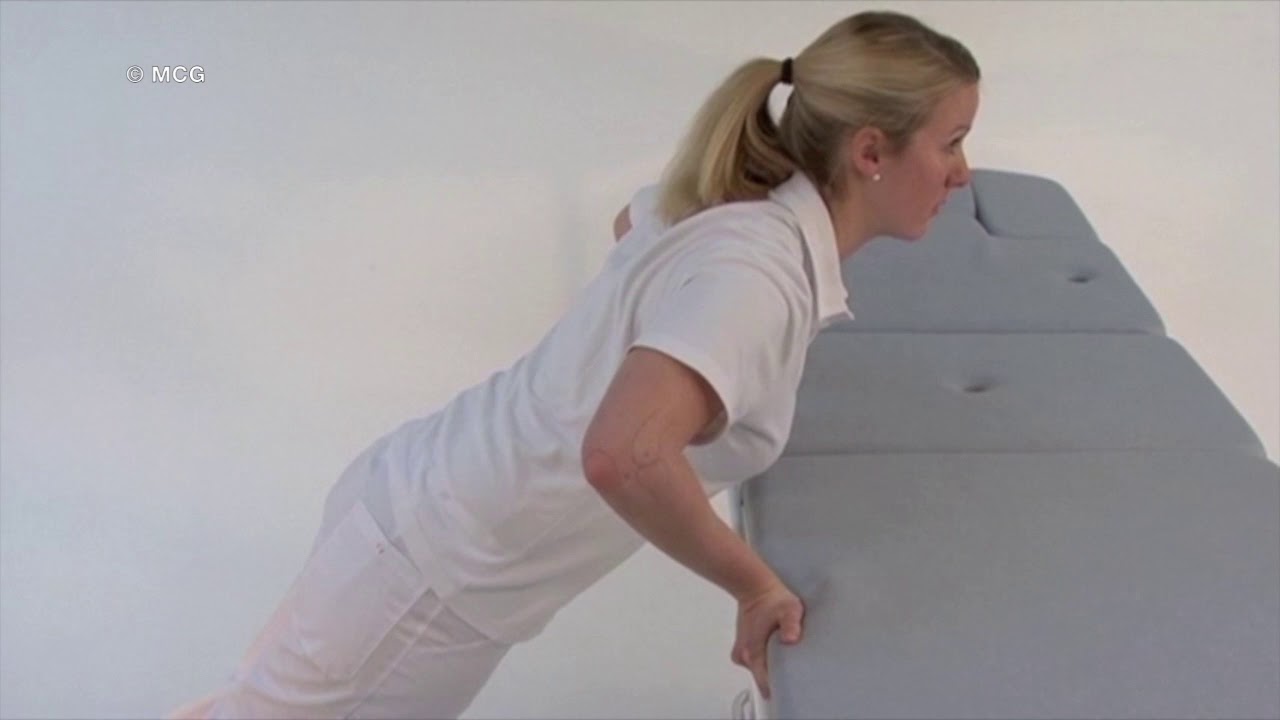
With the following video, we would like to demonstrate a structured clinical exam of the elbow, summarizing the most important maneuvers. We thank Andreas Lutcher and the Shoulders Clinic for their support. The inspection of the soft tissues and bony structures, as well as active and passive range of motion, are tested simultaneously on both arms. Flexion and extension are documented on the supinated arm to avoid an overestimation of the range of motion secondary to internal rotation of the forearm. Pronation and supination are evaluated with the elbows flexed at 90 degrees with the arms at the body. Palpation of the elbow joint focuses on muscle and tendon insertion sites. Both epicondyles, as well as the insertion of the triceps muscle, are assessed for tenderness on palpation. In case of a subsequent arthroscopic intervention, the exact location of the ulnar nerve and a tendency to luxate out of the cubital tunnel have to be documented carefully in the area surrounding the so-called soft spot, which is defined by the electronic radial head and capitulum. Swelling caused by an intra-articular effusion is easiest to locate. The distal biceps tendon can also be palpated easily. A cursory examination of the sensory motor function completes the step of the examination. A structured sequence of functional tests is demonstrated next, focusing on the most common pathologies and deficiencies, namely ulnar instability, posterolateral instability, plica impingement, epicondylitis, ulnar nerve instability, and distal biceps tendon testing. To examine the ulnar collateral ligaments, a variety of tests are available. We will focus on the valgus stress test, the milking maneuver, and the moving valgus stress test. The valgus stress test is performed in the external rotation of the shoulder and with a supinated forearm, as blockage of the olecranon by the fossa olecrani can be avoided in this position. For the same reason, it is important to perform the test not in full extension. During this test, the medial collateral ligament can be palpated in direct comparison to the other arm. Both tenderness and instability can be assessed. The milking maneuver is performed in an upright sitting or standing position by bending the elbow and exerting traction on the patient's thumb. The forearm is maximally supinated, thus specifically straining the internal ligament complex. Pain and increased instability are assessed in comparison to the other arm. The moving valgus stress test is a dynamic evolution of the milking maneuver. A throwing movement is imitated, which is especially helpful in the evaluation of overhead athletes. As in the milking maneuver, the test is performed in an upright standing or sitting position with the shoulder in 90 degrees abduction. The elbow is bent maximally and then extended while exerting constant valgus stress. The test is considered positive if medial elbow pain can be evoked. Typically, pain reaches a maximum between 120 and 70 degrees extension of the arm. The shear angle describes the angle at which pain reaches a maximum, and the shear range is the range of motion perceived as painful. In general, pain arises suddenly, to which the patient reacts with a defensive movement. The objective quantification of posterolateral rotatory instability in a conscious patient is difficult. Even experienced physicians may need to confirm the diagnosis in narcosis or with an arthroscopy. Various active and passive maneuvers are described in the following. The stand-up test is one of the so-called apprehension tests and can be performed easily and actively by the patient. Using axial compression, supination, and valgus stress, subluxation of the radial head is provoked. The apprehension test is positive if the patient avoids the extension of the elbow while raising himself to a standing position out of a chair. The push-up test is a further apprehension test combining axial compression, supination, and valgus of an affected elbow joint. The elbows are flexed at 90 degrees, the forearms maximally supinated, and set further apart than the shoulders. If active extension is avoided, the test is considered positive. The tabletop relocation test is a further development of the two previously described tests and specifically assesses the integrity of the lateral ulnar collateral ligament. The test is composed of three parts. To begin with, the patient practices a press-up maneuver with his arm supported on a table and the affected elbow pointing away from the body. In the second step, the patient exerts pressure on the table by bending his elbow. In the final and third step, the patient repeats the movement while the examiner applies pressure with his thumb to the radial head. During flexion of the elbow without thumb pressure, pain can be reproduced and typically reaches the maximum at 40 degrees flexion. When the maneuver is performed using thumb pressure to stabilize the radial head, patients experience a significant reduction in pain. The posterolateral rotatory apprehension test is a modification of the lateral pivot shift test and is the most sensitive clinical test for posterolateral rotational instability. According to Shauna Driscoll, the first describer of this test, it represents the most sensitive test to diagnose this pathology. The patient is in a supine position. The completely extended elbow is slowly flexed while exerting valgus stress, supination, and axial pressure. If posterolateral instability is present, the radial head will subluxate when the elbow is flexed between 40 and 70 degrees, which is often visualized by skin retraction proximal to the radial head. When the elbow is further flexed and the patient is adequately relaxed, the radial head will reposition itself. The posterolateral rotatory drawer test is a further test to investigate this kind of instability. The positioning of the patient is identical. The elbow is held in the same way as the knee for a Lachman test. One hand grips the patient's upper arm, the other the forearm in maximal supination. In the presence of posterolateral rotatory instability, the forearm can pivot around the intact medial ligaments while performing the drawer movement. In day-to-day practice, examining the elbow using a pincer grip is helpful. The examiner's thumb and forefinger encompass the patient's proximal radius and perform a translational and supinational movement. This patient is a striking example of posterolateral instability following a Homan operation. Arthroscopy confirmed the clinical findings without a doubt. The indication for open reconstruction of the LUCL was given. The osteochondral shear test, also called the grip and grind test, aims to locate osteochondral lesions in the humeroradial compartment. The elbow is actively pronated and supinated while the examiner exerts axial pressure. The test is considered positive if the patient complains of pain in the lateral elbow joint. Apart from the characteristic point tenderness of the tendons inserting at the epicondyles, several provocative maneuvers confirm the diagnosis of epicondylitis. The Cozen's test examines active dorsiflexion of the wrist against resistance. In the presence of radial epicondylitis, the typical pain of the patient will be induced. A loss of strength is also often noted. Active extension of the middle finger against resistance, as described by Maudsley, specifically tests the integrity of the extensor carpi radialis brevis muscle and tendon. Similar to the Cozen's maneuver, flexion of the wrist against resistance will provoke pain if ulnar epicondylitis is present. By testing resistive pronation, the diagnosis of golfer's arm can be confirmed. Additionally, an intra-articular plica can be trapped both in the ventral or dorsal compartment of the elbow joint. To examine the dorsal plica, the elbow is extended and supinated at the same time. In addition, the examiner exerts pressure on the soft spot and so the plica. By performing this test in pronation and flexion, entrapment of a ventral plica can be evoked. This test is a...